
Cervical range of motion (ROM) measures how far the neck joints can move, expressed in degrees across different planes. Healthcare providers use these measurements to assess neck injuries, track rehabilitation progress, and identify movement restrictions that affect daily activities. If you’re standardizing programs across providers, pairing ROM with consistent neck strengthening progressions helps keep care plans objective.
Standard Cervical Movement Measurements
Normal cervical range of motion provides the baseline for evaluating neck function and identifying movement restrictions. These standardized measurements offer reference points for clinical assessment and help distinguish between normal age-related changes and pathological limitations.
The following table provides comprehensive reference values for normal cervical range of motion:
| Movement Type | Normal Range (degrees) | Age Considerations | Active vs Passive Notes
|
|---|---|---|---|
| Cervical Flexion | 40-80° | Decreases 5-10° after age 60 | Active typically 5-10° less than passive |
| Cervical Extension | 50-85° | Decreases 10-15° after age 60 | Minimal active/passive difference |
| Right Lateral Flexion | 45° | Decreases 5° after age 60 | Active typically 5° less than passive |
| Left Lateral Flexion | 45° | Decreases 5° after age 60 | Active typically 5° less than passive |
| Right Rotation | 80-90° | Decreases 10-15° after age 60 | Active typically 10° less than passive |
| Left Rotation | 80-90° | Decreases 10-15° after age 60 | Active typically 10° less than passive |
Key factors affecting normal cervical ROM include:
- Age-related changes: ROM naturally decreases with aging due to joint degeneration and soft tissue changes
- Gender differences: Women typically demonstrate slightly greater ROM than men
- Individual variation: Normal ranges account for anatomical differences between individuals
- Combined movements: Total cervical motion involves complex three-dimensional movement patterns
Cervical ROM Measurement Procedures
Accurate measurement of cervical ROM requires standardized techniques and proper positioning to ensure reliable results. Healthcare providers use various tools and methods to assess neck movement limitations and track treatment progress. For clinics that also track functional capacity over time, consider adding simple endurance testing alongside ROM to better interpret functional change.
Patient Positioning and Setup
Proper patient positioning is crucial for accurate measurements:
- Seated position: Patient sits upright with feet flat on floor
- Shoulder stabilization: Shoulders remain level and relaxed
- Head position: Start in neutral alignment with eyes looking forward
- Spine alignment: Maintain natural cervical and thoracic curves
Manual Measurement Techniques
The following table outlines standardized measurement procedures for each movement plane:
| Movement | Patient Position | Goniometer Placement | Key Technique Points | Common Errors to Avoid
|
|---|---|---|---|---|
| Flexion | Seated, shoulders stabilized | Axis over ear canal, arms along skull and vertical | Move chin toward chest | Shoulder elevation, thoracic flexion |
| Extension | Seated, shoulders stabilized | Axis over ear canal, arms along skull and vertical | Look up while keeping mouth closed | Shoulder retraction, opening mouth |
| Lateral Flexion | Seated, facing forward | Axis over C7 spinous process | Ear toward shoulder, no rotation | Shoulder hiking, trunk side-bending |
| Rotation | Seated, shoulders square | Axis over head vertex, arms along nose and shoulders | Turn head left/right | Shoulder rotation, trunk turning |
If you want to reduce variability between clinicians, objective systems like our Multi-Cervical Unit can support standardized positioning, repeatable testing, and clear documentation.

Best Practices for Accurate Measurement
- Warm-up movements: Allow 2-3 gentle movements before measuring
- End-feel assessment: Note the quality of resistance at end range
- Pain monitoring: Stop if patient reports sharp or severe pain
- Multiple measurements: Take 2-3 measurements and average results
- Documentation: Record measurements consistently using standardized forms
Objective Testing Tools and Techniques
Manual goniometry is the most accessible way to assess cervical ROM, but it carries well-documented limits. Specifically, readings vary between clinicians, single-plane measurements can miss the coupled motion of the cervical spine, and manual measurements are difficult to reproduce consistently from one visit to the next.
When a clinic needs measurements it can compare reliably across a plan of care—and share with referrers and payers—instrumented objective testing helps close that gap.
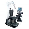
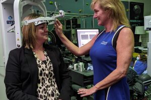
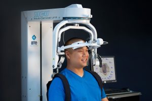
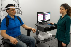
The Multi-Cervical Unit (MCU) is BTE’s dedicated system for objective cervical assessment and rehabilitation. It helps clinicians standardize head and trunk positioning, guide the patient through a repeatable protocol, and capture ROM data without relying on manual goniometer alignment.
Where a goniometer typically records one plane at a time, the MCU evaluation protocol measures cervical ROM across 16 directions—including flexion, extension, and rotation—giving clinicians a more complete picture of where motion is restricted. Few objective systems assess cervical motion this comprehensively, which makes the MCU a useful reference point when standardizing testing across providers.
Because positioning and procedure are standardized, the same test can be repeated by different clinicians in the same clinic with far less inter-rater variability. That consistency is what makes the numbers meaningful when you are tracking progress over time or communicating a patient’s status to a referring physician or case manager.
Reading the MCU ROM Evaluation Report
A central advantage of instrumented testing is the report it produces. The MCU generates an objective ROM evaluation report that presents the patient’s measured motion in each tested direction and compares it against normative values for a healthy population. That comparison makes deficits immediately visible: clinicians and patients can see exactly which directions fall short of the expected range, and by how much, rather than interpreting a column of raw degrees.
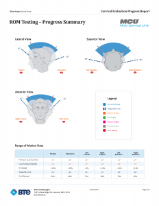
Repeating the evaluation over a course of care turns the same report into a progress-tracking tool. Side-by-side results document measurable change, help clinicians support continuation-of-care and discharge decisions, and give referrers, payers, and employers a clear, objective record of improvement.
For neck pain, whiplash-associated disorders, and other cervical presentations, that documentation is often what distinguishes an objective plan of care from a subjective one.
Clinical Importance and Warning Signs
Cervical ROM limitations can significantly impact daily function and may indicate underlying pathology requiring professional evaluation. Understanding the relationship between movement restrictions and functional disability helps guide appropriate treatment decisions.
Functional Impact of ROM Loss
Limited cervical ROM affects various daily activities:
- Driving safety: Reduced rotation limits ability to check blind spots
- Work performance: Desk work becomes difficult with flexion/extension loss
- Sleep quality: Positioning restrictions can disrupt rest
- Social interaction: Limited rotation affects conversation and eye contact
For teams treating higher-irritability presentations, our guidance on severe neck pain can help clinicians balance symptom management with appropriate measurement timing.
Red Flags Requiring Immediate Attention
The following table provides guidance for identifying serious conditions requiring urgent evaluation:
| Clinical Finding | Severity Level | Recommended Action | Safety Considerations
|
|---|---|---|---|
| Severe pain with minimal movement | Immediate | Stop testing, refer to emergency care | Do not continue ROM assessment |
| Neurological symptoms (numbness, weakness) | Immediate | Refer to specialist immediately | Avoid neck manipulation |
| Recent trauma with ROM loss | Urgent | Medical evaluation within 24 hours | Immobilize until cleared |
| Progressive ROM loss over weeks | Routine | Schedule medical evaluation | Document changes carefully |
| Functional limitations affecting ADLs | Routine | Consider physical therapy referral | Monitor for worsening |
Safety Contraindications for Testing
Avoid ROM testing in the presence of:
- Acute neck trauma: Recent injury requiring medical clearance
- Severe pain: Pain levels above 7/10 that worsen with movement
- Neurological deficits: Weakness, numbness, or tingling in arms
- Vertebral artery insufficiency: Dizziness or nausea with neck movement
- Inflammatory conditions: Active rheumatoid arthritis or infection
Documentation and Progress Tracking
Effective ROM documentation should include:
- Baseline measurements: Initial values for all movement planes
- Pain levels: Associated discomfort during testing
- Functional limitations: Specific activities affected by restrictions
- Treatment response: Changes following intervention
- Goal setting: Target ROM values for rehabilitation
Final Thoughts
Cervical range of motion assessment provides valuable insights into neck function and helps guide treatment decisions for various conditions. Normal ROM values serve as important reference points, but individual variations and age-related changes must be considered during evaluation. Accurate ROM assessment, combined with appropriate clinical judgment, forms the foundation for effective neck injury management and rehabilitation planning.