
Discover how to strategically implement ultrasound machines for physical therapy to improve patient outcomes, streamline workflows, and boost clinic profitability. Expert protocols, device selection criteria, and proven case studies for modern PT practices.
Therapeutic ultrasound remains a valuable modality in physical therapy when used strategically. This guide covers the science behind thermal and non-thermal effects, helps you select the right equipment based on your role (clinic owner, practitioner, or administrator), provides detailed protocols for common conditions, and demonstrates how to integrate ultrasound with other modalities. We’ll show you how to leverage ultrasound therapy not just as a passive treatment, but as a tool that enhances clinical outcomes, improves workflow efficiency, and supports your practice’s growth.
Ultrasound as a Strategic Clinical Asset
If you’re running a modern PT clinic, you know the pressure: deliver superior, evidence-based outcomes while maintaining efficiency and profitability. Every piece of equipment needs to pull its weight, and therapeutic ultrasound is no exception.
While ultrasound therapy has been a staple in physical therapy for decades, many clinics aren’t maximizing its potential. Too often, it’s viewed as just another passive modality—something to fill time or appease patients who expect it. But what if we told you that with the right approach, ultrasound can become a strategic tool that enhances your other interventions, accelerates patient progress, and differentiates your practice?
This guide reframes therapeutic ultrasound as a powerful asset for achieving both clinical and business objectives. We’ll move beyond the basics to explore advanced protocols, integration strategies, and the measurable impact on your practice. Whether you’re a clinic owner seeking to enhance revenue and differentiation, a practitioner committed to evidence-based results, or an administrator focused on standardization and efficiency, you’ll find actionable insights tailored to your needs.
The Core Science Behind Ultrasound Machines
Mechanism of Action, Reimagined
Let’s move past “sound waves create heat” and explore what’s actually happening at the tissue level—and more importantly, how you can leverage these effects strategically.
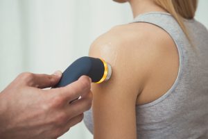
Thermal Effects occur when you use continuous ultrasound waves. The acoustic energy converts to heat, increasing tissue temperature by 1-4°C. This increase enhances blood flow, decreases muscle spasm, and improves tissue extensibility. For your practice, this means you can prep tissues more effectively before manual therapy or exercise, making your hands-on work more efficient and comfortable for patients. Think of it as pre-conditioning the tissue environment for your primary interventions.
Non-Thermal (Mechanical) Effects happen with pulsed ultrasound. Here, cavitation and acoustic streaming create microscopic mechanical forces that influence cellular activity without significant heating. These effects help reduce inflammation, enhance cellular repair, and modify scar tissue formation. This becomes your go-to approach for acute injuries and post-operative patients where heating might be contraindicated but you still need to facilitate healing.
The Critical Role of Frequency: Deep vs. Superficial Targeting
Your ultrasound machine’s frequency setting determines treatment depth, and getting this right is crucial for effectiveness.
1 MHz frequency penetrates 2-5 cm deep, making it ideal for:
- Hip joint capsule restrictions
- Piriformis syndrome
- Shoulder impingement structures
- Deep lumbar paraspinals
3 MHz frequency stays within 1-2 cm of the surface, perfect for:
- Lateral epicondylitis
- Patellar tendinopathy
- Achilles tendon issues
- Carpal tunnel syndrome
Understanding this distinction helps you match your treatment to the anatomical target, ensuring the acoustic energy reaches where it’s needed most.

Choosing Your Ultrasound Device
Let’s elevate the conversation from basic features to strategic capability and return on investment. Your role in the clinic determines what matters most.
For the Clinic Owner/Manager: ROI & Differentiation
When evaluating ultrasound equipment, consider how it fits your broader strategy. Combo units that integrate ultrasound with electrical stimulation maximize your footprint efficiency—crucial in today’s real estate market. Look for devices with pre-set protocols that ensure consistency across your staff while reducing training time.
Consider total cost of ownership beyond the sticker price. A reliable unit with a comprehensive warranty might cost more upfront but saves money long-term through reduced downtime and replacement costs. The ability to provide objective documentation of treatment parameters also supports your value proposition to referring physicians and payers.
For the Clinical Practitioner: Efficacy & Workflow
Your concerns center on clinical effectiveness and daily usability. The ergonomics of the applicator head matter more than you might think—a well-designed transducer reduces hand fatigue during those busy days with back-to-back patients. An intuitive interface saves precious minutes between patients and reduces documentation burden.
Look for features that enhance patient engagement. Visual feedback displays that show treatment progress help patients understand what’s happening, improving buy-in and compliance. This objective feedback also helps you demonstrate progress, much like how BTE’s evaluation systems provide quantifiable data for functional improvements.
For the Hospital/System Administrator: Standardization & Compliance
Your priorities include ensuring consistent care across multiple sites and practitioners. Data and reporting capabilities become essential for tracking usage patterns and outcomes across your system. You need equipment that integrates smoothly with your existing documentation systems and supports quality improvement initiatives.
Ease of onboarding matters when you’re training large numbers of staff. Devices with standardized protocols and clear interfaces reduce training time and minimize variation in application across practitioners.
Evidence-Based Clinical Protocols
Here’s where we bridge the gap between theory and practice. These protocol starting points help you deliver consistent, effective care while always allowing for clinical reasoning based on individual patient presentation.
Lateral Epicondylosis
Clinical Objective: Reduce chronic inflammation and promote tissue remodeling
- Frequency: 3 MHz (superficial target)
- Mode: Pulsed (50% duty cycle) for anti-inflammatory effects
- Intensity: 0.5-1.0 W/cm²
- Duration: 5-7 minutes
- Integration Strategy: Follow with eccentric strengthening exercises and manual soft tissue work to the forearm extensors
Patellar Tendinopathy
Clinical Objective: Increase tissue extensibility and reduce pain before loading exercises
- Frequency: 3 MHz
- Mode: Continuous for thermal effects
- Intensity: 1.0-1.5 W/cm²
- Duration: 5-8 minutes
- Integration Strategy: Immediately follow with progressive loading exercises, starting with isometrics and progressing to eccentrics
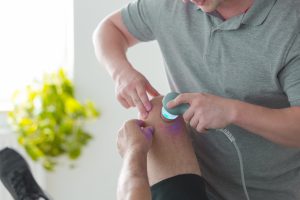
Subacromial Pain
Clinical Objective: Improve capsular mobility and reduce guarding
- Frequency: 1 MHz (deeper penetration needed)
- Mode: Continuous
- Intensity: 1.5-2.0 W/cm²
- Duration: 8-10 minutes
- Integration Strategy: Combine with manual therapy techniques and progress to rotator cuff strengthening
Post-Surgical Scar Management
Clinical Objective: Modify scar tissue formation and improve tissue mobility
- Frequency: 3 MHz
- Mode: Pulsed (20% duty cycle) initially, progressing to continuous
- Intensity: 0.5-1.0 W/cm²
- Duration: 5 minutes
- Integration Strategy: Combine with scar mobilization techniques and progressive stretching

Integrating Ultrasound Machines into PT Practice
The real power of therapeutic ultrasound emerges when you integrate it strategically with other interventions. Think of ultrasound not as a standalone treatment but as a catalyst that enhances your other techniques.
Ultrasound + Manual Therapy
Using thermal ultrasound before manual therapy helps reduce muscle guarding and improves tissue compliance. This preparation makes your manual techniques more effective and comfortable for patients. For example, applying ultrasound to the thoracic paraspinals before mobilization can significantly improve the patient’s tolerance and your ability to achieve the desired movement.
Ultrasound + Therapeutic Exercise
Pain and stiffness often limit exercise quality. By using ultrasound to improve these symptoms temporarily, you enable patients to perform exercises with better form and higher intensity. This enhanced exercise quality accelerates strength and functional gains. Consider how this approach complements equipment like BTE’s PrimusRS, which helps clinicians provide versatile task simulation rehabilitation—ultrasound can prepare tissues for these functional activities.
Ultrasound vs. Electrical Stimulation: When to Choose What
Understanding when to use ultrasound versus electrical stimulation—or both together—elevates your clinical decision-making. Ultrasound excels at tissue heating and mechanical effects at depth, while electrical stimulation provides pain modulation and muscle activation. For comprehensive treatment, consider combination approaches: ultrasound for tissue preparation followed by neuromuscular electrical stimulation for motor re-education, especially in post-surgical cases.
Safety, Contraindications, and Best Practices
The Non-Negotiables
Never compromise on these absolute contraindications:
- Over pacemakers or other implanted electronic devices
- Over reproductive organs or during pregnancy
- Over active infections or malignancies
- Over areas with impaired circulation or sensation
- Over epiphyseal plates in children
Clinical Best Practices
Maintain continuous applicator movement throughout treatment—static application can create hot spots and patient discomfort. Use adequate coupling gel to ensure proper acoustic transmission. Monitor patient response throughout treatment and adjust parameters based on their feedback. Document all treatment parameters for consistency and medical-legal protection.
Conclusion: Make Therapeutic Ultrasound Your Competitive Edge
We’ve explored how strategic implementation of therapeutic ultrasound enhances clinical outcomes, improves workflow efficiency, and provides clear return on investment. By understanding both the science and the strategy, you can transform this traditional modality into a modern clinical asset.
The key lies in viewing ultrasound not as an isolated intervention but as part of your comprehensive treatment approach. When combined with manual therapy, therapeutic exercise, and objective outcome measurement—like that provided by BTE’s evaluation systems—ultrasound becomes a force multiplier for your clinical effectiveness.
Adopting an advanced approach to ultrasound therapy isn’t an expense; it’s an investment in your practice’s future. It helps you deliver better outcomes, differentiate your services, and build stronger relationships with both patients and referral sources.
Ready to elevate your practice with the right rehabilitation and evaluation tools? Explore how BTE’s functional rehabilitation systems can complement your ultrasound therapy protocols and help you provide objective, measurable outcomes that set your clinic apart.
Ultrasound Physical Therapy FAQs:
- How deep can therapeutic ultrasound penetrate into tissues and what structures does it effectively treat?
Therapeutic ultrasound penetrates 2-5 cm deep into tissues, making it most effective for treating superficial to moderate-depth structures. It works best on connective tissues like tendons, ligaments, joint capsules, and fascia. Deeper structures like hip joints or thick muscle groups may require higher frequencies (3 MHz for shallow tissues, 1 MHz for deeper penetration). The energy absorption is greatest in collagen-rich tissues, which is why it’s particularly effective for tendinitis, bursitis, and scar tissue management.
- What’s the difference between continuous and pulsed ultrasound modes, and when should each be used?
Continuous ultrasound delivers constant energy and produces significant thermal effects, making it ideal for chronic conditions where heating tissues is beneficial – such as muscle spasms, joint stiffness, and chronic tendinopathies. Pulsed ultrasound delivers energy in intervals with rest periods, producing primarily mechanical effects with minimal heating. Use pulsed mode for acute injuries, inflammation, or when thermal effects are contraindicated. The duty cycle in pulsed mode is typically 20-50%, allowing tissues to cool between pulses.
- What are the absolute contraindications for ultrasound therapy that clinic staff must never overlook?
Never use ultrasound over: active or suspected malignancies, infected tissues, thrombophlebitis or DVT areas, the carotid sinus, eyes, reproductive organs, or areas with impaired circulation. Avoid use over metal implants (especially near spinal hardware), pacemakers, or other electronic implants. Don’t apply over epiphyseal plates in growing children, pregnant uterus, or areas with impaired sensation where the patient cannot provide feedback about heat buildup. When in doubt, always consult with the supervising therapist or physician.
- How should the ultrasound transducer be moved during treatment to ensure patient safety and effectiveness?
The transducer must be kept in constant motion throughout the entire treatment to prevent hot spots and potential tissue burns. Move the transducer head in overlapping circular or linear patterns at a speed of 1-2 inches per second. Maintain firm, consistent contact with adequate coupling gel – loss of contact creates air gaps that can cause reflection and heating of the transducer. The effective radiating area should overlap by 50% with each pass. If the patient reports any discomfort or unusual sensations, stop immediately and reassess technique.
- What intensity settings should be used for different conditions and tissue types?
For acute conditions or sensitive patients, start with low intensities (0.1-0.5 W/cm²). Chronic conditions typically require moderate intensities (0.5-1.5 W/cm²), while dense scar tissue may need higher settings (1.0-2.0 W/cm²). Never exceed 3.0 W/cm² in clinical practice. Begin conservatively and increase gradually based on patient response. Pulsed mode generally uses higher peak intensities but lower average intensities. Always consider tissue type – more vascular tissues require lower intensities, while dense connective tissues can tolerate higher settings.
- How long should ultrasound treatments last and how frequently should they be administered?
Treatment duration depends on the area size and condition being treated. Small areas (2-3 times the size of the transducer head) typically require 3-5 minutes, while larger areas may need 8-10 minutes. Calculate approximately 1-2 minutes per area equivalent to the transducer head size. Treatments are usually administered 3-5 times per week for acute conditions and 2-3 times per week for chronic conditions. Total treatment courses typically range from 6-12 sessions, with reassessment after 6 treatments to determine continued need. Always document patient response and adjust frequency based on clinical progress.