The Boutonniere Breakdown: A Therapist's Guide to Evaluation, Treatment, and Management of Boutonniere Deformity
Treatment GuidelinesDive into the complexities of managing boutonniere deformity, a common yet challenging finger injury in sports and rheumatoid arthritis patients. This article offers a comprehensive guide on diagnosis, treatment, and practical insights for rehab therapists.
Finger injuries are commonplace in the hand therapy setting. However, in some instances, physical therapists or occupational therapists who don’t specialize in hand disorders may need to manage the care of a finger or hand injury.
One such finger injury is the boutonniere deformity. The name comes from its resemblance to a buttonhole created by the proximal phalanx as it protrudes through the damaged tendon. It commonly occurs as a traumatic injury, most often in basketball and football players. In atraumatic instances, it can develop as a consequence of rheumatoid arthritis.
Regardless of the etiology, boutonniere deformities can be difficult injuries for both patients and clinicians. This is especially true for the therapist who rarely encounters distal upper extremity injuries. In this post, I will provide an overview of the current understanding of how to manage this challenging condition.
Etiology of Boutonniere Deformity
A boutonniere deformity usually results from trauma directly to the digit or a rheumatic condition. The extensor tendon is the chief tissue of concern. The lateral portions of the extensor tendon will split, allowing the phalangeal head to protrude through the new opening created by the disrupted tendon.
Evaluation of Boutonniere Deformity
Often, visual inspection paired with a detailed history will enable you to diagnose the injury. With imaging, the clinician can further assess the damage to the affected structures. Additionally, two special tests can add to your diagnostic confidence: Elson’s test and Boye’s test.

Elson’s Test
Elson’s test is a common physical examination for a boutonniere deformity. To perform the test, the patient hangs their finger over the edge of the table, with the PIP flexed to 90 degrees. Then, the clinician has the patient attempt to extend at their DIP joint. Being unable to extend is a negative finding. Rigidity in the joint during the test is a positive finding, indicating that the central slip of the extensor tendon is disrupted.
Boye’s Test
During Boye’s test, the patient’s PIP is held in extension and the patient will actively flex at the DIP joint. Inability to perform flexion at the DIP joint in this position is a positive finding, indicative of an extensor tendon disruption. However, it’s important to note that Boye’s test is usually only positive in advanced injuries or those that have been present for quite some time.
Differential Diagnosis of Boutonniere Deformity
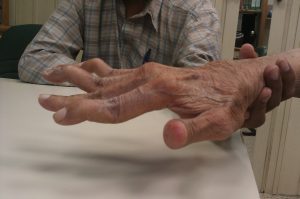
Most cases of boutonniere deformity are fairly straightforward. However, clinicians who lack experience with hand and digit injuries should be careful to ensure that they have correctly diagnosed the issue. Up next we’ll discuss four other conditions that may present similarly to a boutonniere deformity and how you can tell the difference. The image below shows a boutonniere deformity on two fingers of a patient with rheumatoid arthritis.
PIP Joint Dislocation
At first glance, a PIP dislocation looks very similar to the condition of interest. That being said, PIP joint dislocations will often present with more marked symptoms when compared to the boutonniere deformity.
Burn-related Skin Contracture
In burn rehabilitation, skin contractures are a common issue as they can limit joint movement. If a burn-related skin contracture limits joint movement in a similar pattern, you might be tempted toward a diagnosis of boutonniere deformity. Again, this should be easy to rule out with a thorough history and examining the skin integrity.
Swan Neck Deformity
Much like many boutonniere deformities, swan neck deformities often occur due to trauma. In contrast, the swan neck deformity will show hyperextension of the PIP joint and flexion of the DIP joint.
Mallet Finger
At first glance, a mallet finger can resemble a swan neck deformity. However, mallet finger only involves the DIP in an extension lag, the PIP is not involved. Patients with mallet finger will be unable to extend the finger at the DIP joint.
Conservative Treatment of Boutonniere Deformity
Many cases of boutonniere deformity require surgery, especially in traumatic injuries. But in cases where surgery is contraindicated or otherwise not a viable option, a physical therapist or occupational therapist has a few options at their disposal.
Splinting
Splinting is a go-to nonsurgical treatment option for a boutonniere deformity. Many protocols will require the patients to splint the affected digit for 3-6 weeks, wearing the splint at night following the initial immobilization period.
Exercise
Regardless of whether or not surgery is indicated, most patients will complete ROM and strengthening exercise for the affected digit and hand. Maintaining range of motion is of paramount importance for this condition.
Medication
In cases related to rheumatoid arthritis, patients may be prescribed a variety of medications to help manage a boutonniere deformity. These include DMARDS, pain relievers, and a variety of other options depending on the patient’s individual symptoms and needs.
Prognosis
Naturally, prognosis will depend largely on the severity of the injury and other patient-specific factors. However, many patients with more severe boutonniere deformities will not recover full function or ROM in the affected digit.
The clinician should strive to help the patient maintain as much ROM and strength as possible and minimize any complications. A return to athletic competition may require the use of a brace, tape, or splint in perpetuity.
Conclusion
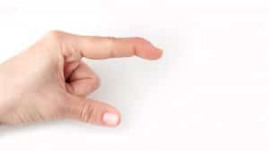
A boutonniere deformity is a relatively common digit injury that results from damage to the central slip of the extensor tendon of a finger. This injury often leads to a characteristic posturing of the involved digit, in which the patient maintains PIP flexion and DIP extension, as if they are “pinching” something.
Managing this condition can be tricky, and many advanced cases will require surgery, along with other conservative methods, such as splinting. Unfortunately, boutonniere deformity may result in continued functional deficits along with decreased ROM in the involved digit. Rehab professionals should be familiar with this issue, as it is observed in both athletic populations and in those with rheumatoid arthritis.
Bennett Richardson, PT, DPT, CSCS is a Physical Therapist and writer. He is the owner of Richardson PT LLC, a mobile, cash-based physical therapy service out of Pittsburgh, PA. Ben is passionate about many health-related topics including weight loss and athletic performance. To get in touch with Ben, visit www.richardsonpt.com